Lissencephaly
What is Smooth Brain or Lissencephaly?
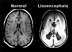
Lissencephaly, from the Greek words lissos for smooth and cephale for head. Usually, a normal brain is folded upon itself with ridges (gyri) and fissures (sulci). These folds begin to develop in the last trimester of pregnancy (at 26 to 28 weeks gestation) and give the brain more surface area, allowing for the development of more neurons (nerve cells) and for more communication between the neurons.
Only about 10 to 40 per million babies are born with some level of lissencephaly, so this is a fairly rare condition. The abnormalities in these babies’ brains can be limited or can be very pronounced, depending on the severity of the lissencephaly.
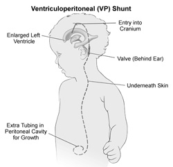
Severely affected babies will have profound developmental delays, possibly including decreased muscle tone (due to inadequate nerve stimulation of the muscles) and/or decreased ability to feed (and hence grow). They may also develop hydrocephalus, sometimes called fluid on the brain, where too much of the normal fluid around the brain (cerebrospinal fluid) accumulates which can cause increased pressure inside the baby’s skull. Hydrocephalus may require treatment with a VP shunt, a tube that goes from the brain through a hole placed in the skull and then under the skin all the way to the abdomen, allowing the excess fluid to drain. Babies with lissencephaly may also develop seizures, which are sometimes difficult to treat even with aggressive and multiple anti-seizure medications.
Lissencephaly is usually due to a genetic defect in chromosome 17, causing a problem with the protein reelin which regulates the migration of neurons and the positioning of the fetus’ developing brain. Other genetic defects, including one linked to the X chromosome, have also been implicated in some cases. There are several genetic syndromes that have a constellation of birth defects which have lissencephaly as one of the conditions. Some babies born with lissencephaly also have abnormal fingers, toes or even hand development.
Other conditions that affect the fetus’ brain development, such as fetal viral illnesses or conditions that decrease blood flow to the fetus’ developing brain, might also be causes of lissencephaly in certain cases.
Babies who have developmental delay and/or seizures will often be evaluated by an ultrasound, MRI or CT scan of their brain, and if lissencephaly is present this is usually how it is diagnosed. Since, as noted above, a fetus’ brain folds begin to be developed in the last trimester of pregnancy (at 26 to 28 weeks gestation), lissencephaly cannot reliably be seen on prenatal ultrasound unless one is done late in the pregnancy.
The prognosis for babies with lissencephaly is poor, with the specific prognosis for a given child depending on the severity of the brain malformation. Many babies with lissencephaly do not live past their first 2 years of life, often dying of respiratory conditions (such as pneumonia) due to the poorly developed breathing muscles. Some lissencephaly children with severe malformations may live longer, even though they may not develop beyond a 3- to 6-month-old level. Children with less severe malformations may have near normal intelligence and development.
There are no treatments for lissencephaly however associated conditions, such as hydrocephalus or seizures, can be treated, and supportive care for this severe disorder can be offered to the infant. Support for the family of the affected infant is also important.
Source: Dr. Jeff Hersh, Ph.D., M.D. (Natick Bulletin)


Recent Comments